As a child, Todd P. Margolis didn’t think medicine would become his career.
“I wanted to be a scientist,” recalled Margolis, who grew up in Los Angeles. “The concept of being a practicing physician really wasn’t on my radar screen.”
His parents taught their kids to want to make a difference in the world. His brother, Peter, grew up to be an internist in Southern California, and his sister, Ellen, now runs a Meals on Wheels program, which provides meals for older adults. In college, Margolis was aiming for a career in research when his friends talked him into medical school. Although he wasn’t sure about working with patients, he was convinced he’d have more options and a better chance to make a difference if he earned a medical degree and a doctorate.
After graduating from Stanford with a bachelor’s degree in biology, Margolis attended the University of California, San Francisco, where he earned a doctorate in neuroscience. Although he was working toward his medical degree, his career path hadn’t yet been decided.
“I was doing general medical work at the Haight-Ashbury free clinic when friends suggested I think about ophthalmology,” said Margolis, the Alan A. and Edith L. Wolff Distinguished Professor and head of the Department of Ophthalmology and Visual Sciences at Washington University School of Medicine in St. Louis.
It fit well with his interest in neuroscience, and there were plenty of opportunities for research. In addition, eye surgery was similar to the microdissection he’d done in his graduate research, and it turned out to be a good fit.
Going viral
Margolis stayed at UCSF for his residency and a fellowship, focusing on research work with herpes simplex virus in the laboratory of his graduate adviser, Jennifer LaVail, PhD. The herpes virus causes cold sores, but while it typically appears as a rash elsewhere on the body, in the eye it can cause disease that leads to significant damage and vision loss.
“I was seeing patients with herpes infections of the eye and trying to find answers to basic questions about mechanisms, but there was no data,” he recalled. “So in my spare time on weekends and evenings, I went back to my thesis adviser’s lab and did research to obtain answers to some of the questions relevant to patients I was seeing.”
His work eventually led him to UCLA, where he did a postdoctoral fellowship in viral pathogenesis. When he later returned to San Francisco, the city had become ground zero for the AIDS epidemic, and his expertise as a virologist was very much in demand.
Thousands got sick, became immunocompromised and developed opportunistic infections. Margolis went to San Francisco General Hospital to run the AIDS eye clinic. There, he quickly learned about diagnosing and treating viral infections in the eye, particularly infections that caused cytomegalovirus (CMV) retinitis, an inflammation of the retina that can lead to blindness.
Soon, his laboratory was developing molecular tests for opportunistic eye infections in AIDS patients and figuring out what was causing the infections and how to treat them. As the epidemic raged, Margolis received samples from all over the country with requests for diagnosis and suggested treatments.
There’s an app for that
The introduction of anti-retroviral therapy in the mid-1990s prevented AIDS patients from dying in such great numbers. As a result, the demand for Margolis’ work on the diagnosis and treatment of opportunistic eye infections in patients with HIV also dropped off.
“We had an enormous amount of information about diagnosing and managing patients with these problems, and we still wanted to apply it,” he said.
So as the new millennium was beginning, Margolis and his team looked overseas.

He had trained fellows from around the world, and several from Thailand suggested he go there to help out. There, he discovered that the physicians didn’t really need help diagnosing and treating HIV patients with opportunistic eye infections. Their main need was screening.
“I came in quite naively and asked what we could do to help in terms of managing these diseases,” he recalled. “The big problem was that only about six ophthalmologists in the whole country were willing to see patients with HIV, and there were about 1 million HIV patients who needed to be screened for CMV retinitis and other eye infections.”
The need for such massive screening gave Margolis an idea he has been honing for several years.
“I thought if we could take pictures of the back of the eye with a cellphone camera, we could send the images to an expert who could read them from a distance,” he explained. “Ultimately, I borrowed someone’s iPhone and literally stuck it right in front of an eye. I illuminated the eye with a light, and I could see the optic nerve.”
Because cellular networks tend to be better developed than computer networks in many parts of the world, using cellphone cameras to screen patients cheaply, and without a doctor on hand, made more sense than trying to feed pictures over the Internet.
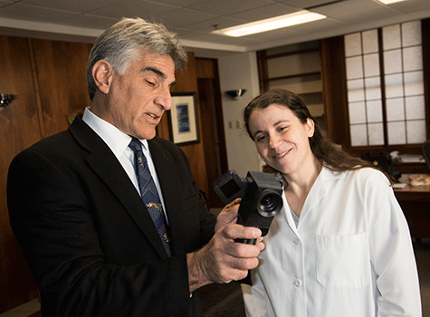
Margolis made prototypes of a fundus camera — used to photograph the interior surface of the eye — to connect with an iPhone. He then collaborated with Daniel Fletcher, PhD, a biomedical engineer at the University of California, Berkeley, who was using cellphone imaging to look at blood smears for malaria in the developing world. The first version they developed together now sits on a shelf in Margolis’ office.
To test their idea, they used a very expensive fundus camera to prove it was possible to screen patients without a visit to a doctor’s office. That led to a series of studies involving the cellphone-based camera and reading images it collected.
A company called Eyenuk has since designed image-recognition software for their camera to recognize the characteristic pattern of CMV retinitis without anyone having to read the image. The company also is working on a method of screening for diabetic retinopathy, a problem that is increasing in the developing world as our fast-food, Western diet spreads to other countries and the obesity epidemic follows.
“In India, for example, the number of patients with diabetes has exploded in the past 10 years,” Margolis explained. “They don’t have enough doctors to take care of their cataract problem, let alone the growing problem with diabetic retinopathy. It’s important to screen those patients to prevent an epidemic of vision loss.”
Screening for diabetic retinopathy is where tools like Margolis’ cellphone-based camera also might come in handy at home.
“We have some amazing tools for managing patients with diabetic retinopathy that decrease their visual problems tremendously, but first you have to identify the patients who need to be treated,” he said. “In the United States, only 50 percent of people with diabetes who are supposed to get eye exams actually get them. But if we set up a camera at the corner pharmacy or local health clinic right next to the blood pressure machine, we could screen a lot more patients for diabetic eye disease when they come to pick up their oral medicines or insulin.”
With the family for dinner
Margolis arrived in St. Louis to head the ophthalmology department earlier this year. Despite the demands of his job, he is the cook at home. While raising his family in California, he always made a point to be home in time for dinner. Making dinner serves as a sort of therapy for him.
“It’s like being in the lab,” he said. “You get to try new things, but then you get to eat them. For me, going to the grocery store or farmers’ market is an adventure, hoping to find something new that I can play with.”
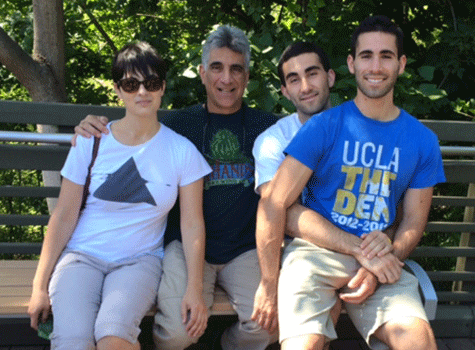
Margolis has three grown children. His daughter, Jessie, 24, just completed her master’s degree in biology at the University of California, San Diego. Mathew, 23, is a first-year medical student at Washington University. Josh, 20, is a business major at the University of Oregon.
And there will be another Margolis soon. About a year ago, he remarried. His wife, Kathleen Apakupakul, also is a scientist. She helped him move his laboratory from San Francisco to St. Louis. And it was her eye that first helped Margolis determine it was possible to use an iPhone to screen for retinal problems. The two are expecting a child any day.
“I loved having a family. I still do,” he said. “And I’m really excited to have another child around the house.”
