A workman, the saying goes, is only as good as his tools, and that is nowhere more true than in science, where researchers are often working blindly until technology catches up and allows them to see what they once could only infer.
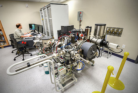
This is why scientists at Washington University in St. Louis are excited that the U.S. Department of Energy (DOE) has awarded $2.4 million to David Fike, PhD, associate professor of earth and planetary sciences, and a team of three other researchers from the International Center for Advanced Renewable Energy and Sustainability (I-CARES) to adapt a powerful chemical microscope called the 7F-GEO SIMS for biological samples.
The 7F-GEO SIMS was purchased in December 2013 with funds from the National Science Foundation’s Major Research Instrumentation program. The microscope is housed in the Grossman Family SIMS Laboratory, which was funded by a gift from Jerrold and Marsha Grossman and their son, Washington University alumnus Matthew Grossman and his wife, Katalin Frisch.
“Because of its high precision, the GEO SIMS is ideal for examining geological samples,” Fike said, “but it doesn’t currently have the spatial resolution to map the internal chemistry of biological cells.”
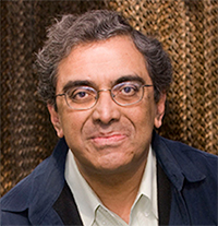
“I’ve always been interested in using it for biology,” he said, “so when Himadri Pakrasi emailed me a call for proposals from the DOE for instruments for biological imaging, a group of us got together and came up with a proposal to upgrade the GEO SIMS.”
Pakrasi, PhD, the Myron and Sonya Glassberg/Albert and Blanche Greensfelder Distinguished University Professor and director of I-CARES, is a co-investigator on the grant.
“We plan to put a novel ion source on the SIMs that has a much smaller spot size so we can make higher resolution images, and also a new detector system on the back end that allows us to collect large field of view images more quickly,” said Fike, the principal investigator.
The instruments now available to Washington University researchers raster a beam across the sample, a process that is very slow. Once the SIMS is upgraded, it will be able to project the entire field of view onto the new detector and record it all at once, Fike said.
It’s a bit like the difference between an old TV painting an image on a cathode ray tube screen line-by-line and a camera instantaneously exposing film.
In its new operating mode, the SIMS will be able to make high-resolution maps of the elements and isotopes (variant forms of elements) within many microbes simultaneously and, by taking repeated images, reconstruct the chemistry of a microbe’s interior in three dimensions.
The group hopes this new-found ability to see inside cells that are only micrometers in diameter will allow them to break stalemates in their work with microbes that are promising candidates for biofuel or bioenergy production.
For example
The three biologists who are co-investigators on the grant each work with a different microbial system and face a different sort of imaging challenge. “They’re going to test drive the instrument and make sure it can do everything we want it to do,” Fike said.
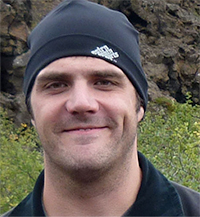
The three are: Pakrasi; Arpita Bose, PhD, assistant professor of biology; and Alex Bradley, PhD, assistant professor of earth and planetary sciences. With the inclusion of Fike, the team was brought together through I-CARES.

Pakrasi has a long list of questions he’d like to answer, but one in particular illustrates the potential of the new instrument. It has to do with a green bacterium called Cyanothece, some species of which can both photosynthesize (fix carbon) and convert nitrogen to a biologically useful form (fix nitrogen).
They’re not supposed to be able to do both at once because the oxygen produced by photosynthesis poisons the enzyme needed to fix nitrogen. So usually the microbes photosynthesize during the day and fix nitrogen at night.
However, Pakrasi’s group has observed that Cyanothece cultures will grow even if they are continuously exposed to light, which implies they have a mechanism to fix nitrogen and perform photosynthesis at the same time.
Are there separate populations of nitrogen-fixing and photosynthesizing cells, he wonders? And do they communicate with one another to decide who does what?
The BIO SIMs will be ideally suited to answer this question, Fike said. Other instruments can only image one or a few cells in detail. “But the BIO SIMS will have a large field of view and will rapidly examine a huge number of cells to figure out who’s fixing nitrogen and who’s not and whether and how the cells are communicating with one another.”
Once Fike and his colleagues have finished honing it, the BIO SIMS will be the sharpest tool in the shed.