Researchers at the University and elsewhere recently helped the St. Louis Science Center probe the mysteries of a baby mummy. The mummy, part of the Science Center’s collection of artifacts for two decades, went on permanent public display on March 15 in conjunction with the arrival of an IMAX film on mummies.
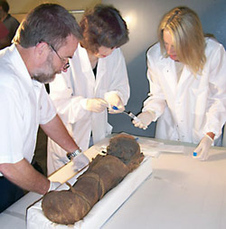
The mummy was donated in 1985 to the Science Center by the niece of a St. Louis dentist. Her uncle purchased it during a trip to the Middle East sometime around the beginning of the 20th century. Parts of the mummy’s wrappings had been cut away at some point, revealing the baby’s facial features, neck and chest. To learn more without inflicting further damage, Science Center officials turned to the Mallinckrodt Institute of Radiology at the School of Medicine.
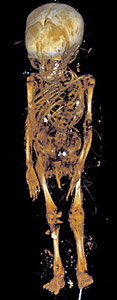
Charles Hildebolt, Ph.D., associate professor of radiology, led a team that used X-ray computed tomography (CT) scanning to create three-dimensional images of the mummy.
“With these images, it’s not only possible to virtually remove the mummy’s wrappings, we can also peer inside the body,” Hildebolt explained.
Using data from the CT scans, Hildebolt and colleagues determined that the mummy was a boy. To assess his age at time of death, they looked at the development of his teeth, closure of the bones in his skull and the formation of his hands, all of which suggested that the boy was seven to eight months old when he died.
The scans showed extensive evidence of procedures often used in mummification. The boy’s brain, for example, had been removed through a break in his ethmoid bone, which separates the nasal cavity from the brain. His internal organs, including the liver, stomach, lung and intestines were removed from a cut in the upper left side of the abdomen.
“Generally the heart was not removed during mummification because the Egyptians thought it was the seat of the soul,” Hildebolt said. “They believed the souls of the dead were able to reanimate their bodies in the hereafter.”
Researchers noted that the child’s head seems large and slightly flattened. The former suggests but does not prove that the baby may have suffered from hydrocephalus, an enlargement of the head caused by fluid buildup in the skull. The latter is a common and usually harmless condition known as plagiocephaly that may have resulted from the baby being kept in one position after birth.
The scans produced no definitive evidence of what caused the boy’s early demise.
“Juvenile mortality was high at that point in world history, and it’s possible that whatever killed him left no trace on his bones,” Hildebolt said.
The baby was wrapped in an outer burial shroud and at least eight layers of linen bandages tied in place with linen bands. To determine what time period the mummy came from, scientists sent a postage-stamp-sized sample of the linen bandages to a radiocarbon-dating firm in Florida. The firm’s analysis found a 95 percent probability that the baby’s brief life occurred sometime between 40 B.C. and 130 A.D.
“This places the baby in the Ptolemaic or Greek period of Egyptian history,” Hildebolt said. “He likely lived during the time when Egypt was a province of the Roman Empire.”
The CT scans also revealed a number of amulets placed throughout the body. According to Hildebolt, these charms and the superior quality of the mummification techniques suggest that the baby was probably a child of a middle-class family that could afford to pay for such services. Historians believe that when Egypt was a Roman province, both Egyptians and Romans may have had family members mummified.
Hildebolt obtained four tissue samples from different areas of the mummy and gave them to colleague Anne Bowcock, Ph.D., professor of genetics, of medicine and of pediatrics for analysis. Bowcock’s group was able to extract DNA from three of the samples and to sequence DNA from the mitochondria, energy-making structures in human cells thought to be descended from symbiotic bacteria. Mitochondria are passed on through the mother, and traits in the DNA suggested the baby’s mother might have been European and possibly Greek or Roman. Bowcock plans further analysis to see if she can determine the geographic origin of the father by looking at the mummy’s Y chromosome.
Research team members in alphabetical order by institution:
American University in Cairo Salima Ikran, PhD (Egyptologist/Mummy Specialist)
Florida State University Dean Falk, PhD (Anthropologist)
Metropolitan Museum of Art Emilia Cortes (Conservator)
Saint Louis Science Center Melinda Frillman, Collections Manager
Washington University School of Medicine Charles F. Hildebolt, DDS, PhD (Dentist, Anthropolgist) Kirk Smith, BS (Senior Research Engineer) Anne Bowcock, PhD (Geneticist) Li Cao, PhD (Geneticist) Steven Don, MD (Pediatric Radiologist)